The advances in technology have enabled the development of powerful microscopes to view the samples at a nanometer level and thus were born the electron microscopes.
What is Electron Microscope
As the name suggests, an electron microscope utilizes a beam of accelerated electron to illuminate the sample to be viewed.
Built first in 1931, the electron microscope was built by a German academician and engineer Ernst Ruska and the same principles continue to be the basic structures for modern day electron microscopes.
Electron microscopy is a modern-day technique that helps obtain high resolution images of non-biological and biological specimens.
Types of Electron microscopes
- Transmission Electron Microscope
- Scanning Electron Microscope
- Reflection Electron Microscope
- Scanning Transmission Electron Microscope
1. Transmission Electron Microscope (TEM):
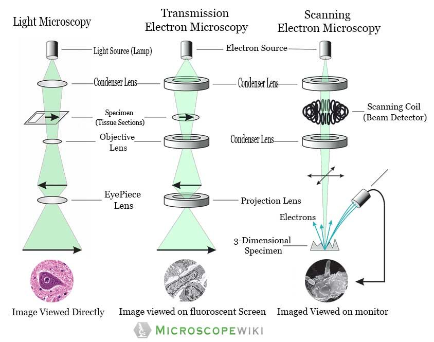
- A transmission electron microscope employs an electron beam produced at high voltage to brighten the specimen and produce an image to be viewed
- The working principle of this microscope is that the electrons pass through the exhibit and create a projection image of the specimen
- TEM is typically used to view thin samples such as molecules, tissue sections etc.
- TEM is similar to a compound microscope but it used to achieve a very degree of magnification thereby allowing the observation of specimens at a nanometer level.
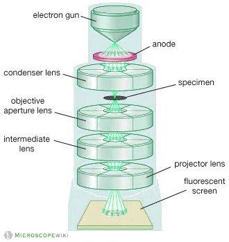
Image : Transmission Electron Microscope (TEM) diagram
2. Scanning Electron Microscope (SEM):
- An SEM creates magnified images of the specimen by probing along a rectangular area of the specimen with a focused electron beam. This process is called the raster scanning.
- It is called a scanning electron microscope because the image is formed by scanning the surface of the specimen in a raster pattern using a focused electron beam.
- SEM relies on the secondary emission of electrons from the surface of the specimen to achieve magnified image to be viewed
- The major advantage of a SEM over TEM is that it can produce detailed image of the whole organisms and surfaces of the cells
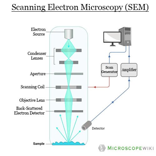
Image 2 a : Scanning Electron Microscope (SEM) Diagram
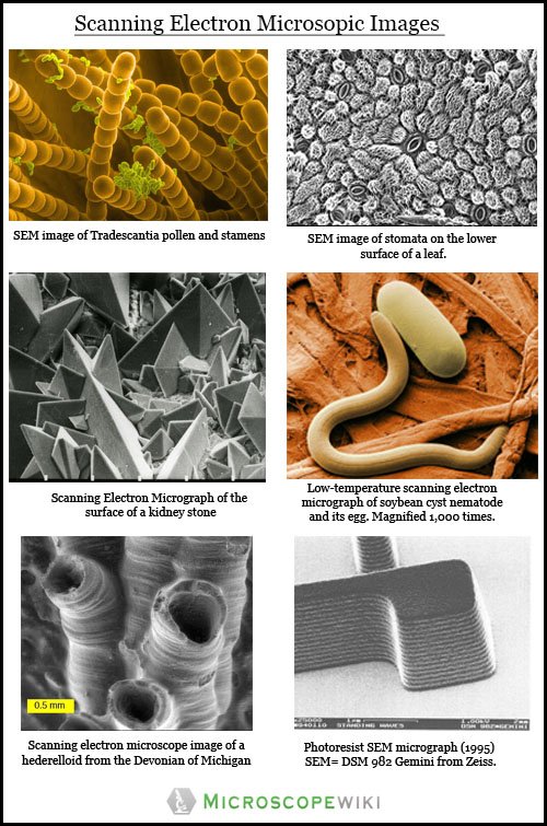
Image 2 b : Scanning Electron Microscope (SEM) image
3. Reflection Electron Microscope (REM):
- As with the other electron microscopes, a beam of electrons is used to create the image. However, this microscope uses neither scattered electrons nor transmitted electrons to create the image. It employs a reflected ray of electrons that have been scattered elastically .
- High-energy electrons are incident at glancing angles to the surface and reflected electrons are used to form an REM image, a Reflection High Energy Electron Diffraction pattern (RHEED), and a Reflection High-Energy Loss Spectrum (REELS)
4. Scanning Transmission Electron Microscope (STEM):
- As the name suggests, STEM is a hybrid approach that employs a combination of transmission electron microscopy and scanning electron microscopy to achieve a better quality of images required for observing a sample.
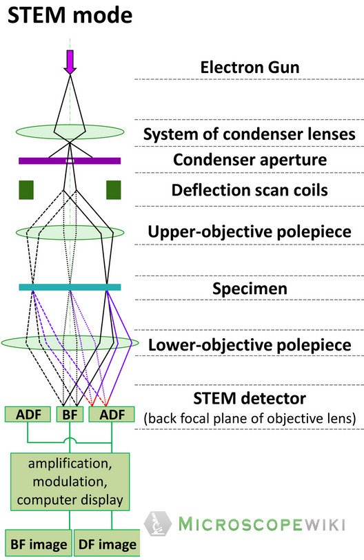
Image 3 : Scanning Transmission Electron Microscope (STEM) diagram
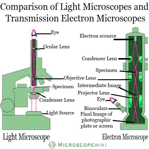
Comparison of Light microscope and Transmission electron microscope
Difference between – Light microscopy, Transmission electron microscopy and Scanning electron Microscopy
Parts of a Transmission Electron Microscope
There are four major components in a transmission electron microscope. They are
- Electron Gun
- Electromagnetic Lens
- Specimen Holder
- Image viewer and recording mechanism
- Electron Gun: This is the part that generates the electrons. The electron gun houses a tungsten filament that is heated to produce electrons
- Electromagnetic Lens: The electromagnetic lens are a combination of three types of lenses which aid in creating an image of the specimen:
- Condenser lens: This lens focuses the electron beam on to the specimen. There is also a second condenser lens that focuses the electrons into a thin beam
- Objective lens: The electron beam that comes out of the specimen and passes through the second set of magnetic lenses is usually termed as the objective lens and this lens produces the intermediate magnified image of the specimen.
- Ocular (projector) lens: The final image of the further magnified image is produced by the projector lens which are another set of magnetic lenses that create higher magnification of images, all the while maintaining excellent level of resolution and detail
- Specimen Holder: This is very thin carbon film or a collodion that is held in place by a metal grid
- Image viewer and recording mechanism: The final magnified image of the specimen appears on the fluorescent screen. There is also a camera right below the screen that is used to record the image.
Applications
- Scientific Research: Electron microscopes find extensive usage in nanotechnology centres, universities, and research labs. The electron microscopes help researchers, students, and scientists to study specimens, both biological and non-biological at a nanoscale to be able to draw deep and meaningful insights
- Natural Resources: Electron microscopy is widely used in characterization and analysis of organic materials making them indispensable for mining companies in search of natural resources. Also, oil and gas companies also use these microscopes in their exploration of oil and gas reserves
- Industry: Electron microscopy is also used widely for industrial applications to assist them in improving efficiencies in manufacturing processes and in developing new and better products. Electronics manufacturing industry, apparel, automotive, pharmaceutical, and aeronautical industries among others use electron microscopes
- Forensic Science: In the process of unravelling a criminal case, electron microscopes find application as they help in analysis of the evidence at a nanoscale. For instance, analysis of samples like hair, residue from gunshot, cloth fibres, biological evidence material like skin, tissue etc. using an electron microscope yields better results.
Advantages
- High Magnification: Modern-day electron microscopes have an extremely high magnification power and they can magnify a specimen up to 500,000 times. The higher magnitude of magnification allows us to see the minute details.
- High Resolution: Apart from the magnification, electron microscopes have superior resolution power and they can distinguish the features, details and properties of a specimen at a nanoscale level thereby enabling better research and better outcomes.
- Compatibility: Electron microscopy is compatible with other technologies and allows for better analysis. For instance, users can see colour images, three dimensional imaging and real-time analysis of specimens
- Versatility: An electron microscope can be used to observe and analyse just about any kind of specimen both inorganic and organic making them indispensable in the fields of industry, biomedical research, forensics among others
Disadvantages
- No colour images: Electron microscopes only create black and white images and they need to be colorized falsely
- Cost: Electron microscopes are very specialized equipment and are very costly to acquire and maintain. As many projects have limited budgets, acquiring and maintaining an electron microscope can be detrimental to their efforts
- Can’t observe live specimens: The very fact that the specimens need to be observed in vacuum renders the observation of live specimens useless as it is near impossible to stay alive in vacuum.
- Specialized knowledge required: The operation of an electron microscope is complex and requires specialized knowledge and training t be able to operate it and maintain it.
Electron Microscope price range
The cost of a new electron microscope ranges between $80,000 to $10,000,000 and above depending on the customizations, configurations, resolution, components, and brand value. The type of electron microscope also decides the price of the microscope because of the various uses the microscope has and also on the components used in them.
The common types of electron microscopes that find widespread usage are Scanning Electron Microscopes (SEM), Focussed ion beam electron microscope (FIB) and Transmission Electron Microscope (TEM).
The table below gives you a fair bit of idea of prices about used as well as new electron microscopes.
| Model | Type | Used Price | Estimated Price (New) |
| FEI Tecnai F20 | TEM | $395,000 | $592,500 |
| FEI Tecnai G2 F30 Twin | TEM | $395,000 | $592,500 |
| FEI Altura 810 | SEM | $350,000 | $525,000 |
| Philips CM 200F TWIN | TEM | $300,000 | $450,000 |
| FEI Tecnai 20 TWIN | TEM | $300,000 | $450,000 |
| Hitachi SU-70 | SEM | $275,000 | $412,500 |
| Hitachi 5500 | SEM | $250,000 | $375,000 |
| FEI Strata 210 | SEM | $250,000 | $375,000 |
| JEOL 1200ex II | TEM | $250,000 | $375,000 |
| FEI FIB 800-M | FIB | $250,000 | $375,000 |
| Hitachi 4700-II | SEM | $225,000 | $337,500 |
| FEI Tecnai 12 | TEM | $225,000 | $337,500 |
| FEI Quanta 200 | SEM | $200,000 | $300,000 |
| FEI FIB 200-S | FIB | $200,000 | $300,000 |
| Hitachi 5200 | SEM | $175,000 | $262,500 |
| Hitachi 4700-I | SEM | $175,000 | $262,500 |
| FEI CM200 | TEM | $175,000 | $262,500 |
| Philips CM 120 BT | TEM | $175,000 | $262,500 |
| FEI FIB 200-M | FIB | $175,000 | $262,500 |
| Philips CM12 | TEM | $161,000 | $241,500 |
| FEI XL40 | SEM | $155,000 | $232,500 |
| FEI XL30 | SEM | $150,000 | $225,000 |
| ZEISS EVO 50 | SEM | $150,000 | $225,000 |
| JEOL 1011 | TEM | $150,000 | $225,000 |
| Philips CM 120 | TEM | $150,000 | $225,000 |
| JEOL 1200ex I | TEM | $150,000 | $225,000 |
| JEOL JEM-2011 | TEM | $150,000 | $225,000 |
| Philips CM 100 | TEM | $150,000 | $225,000 |
| FEI FIB 200-P | FIB | $150,000 | $225,000 |
| FEI Morgagni | TEM | $135,000 | $202,500 |
| FEI Quanta 400 | SEM | $130,000 | $195,000 |
| Philips CM10 | TEM | $125,000 | $187,500 |
| Philips EM 208 | TEM | $125,000 | $187,500 |
| Hitachi 7000 | TEM | $105,000 | $157,500 |
| Zeiss EVO 40 | SEM | $95,000 | $142,500 |
| Hitachi 3000N | SEM | $85,000 | $127,500 |
Frequently Asked Questions (FAQ)
Q1. How powerful is an electron microscope?
Ans: An electron microscope can magnify a specimen between 1 and 50 million times depending on the need of the user and the specimen that is to be observed.
Q2. What is the smallest object that can be seen with an electron microscope?
Ans: The electron microscope can observe and analyse objects at a nanometer scale. For instance, the electron microscope allows the observation of a specimen at an atomic level.
Q3. Why is the electron microscope so big in size?
Ans: An electron microscope has an evacuated column that is vacuum sealed and houses a cathode, anode, condenser magnet, scatter aperture, specimen chamber, objective lens, fluorescent screen, photographic plate and its transport machinery. This is the reason why the microscope is bulky in size.
Q4. What is the difference between a light microscope and electron microscope?
Ans: A light microscope has a low resolving power (0.25µm to 0.3µm) while the electron microscope has a resolution power about 250 times higher than the light microscope at about 0.001µm. Similarly, a light microscope has a magnification of 500X to 1500x while the electron microscope has a much higher magnification of 100,000X to 300,000X.
References:
- https://cores.research.asu.edu/sites/default/files/2018-10/reflection_electron_microscopy.pdf
- https://www.fei.com/introduction-to-electron-microscopy/sem/#gsc.tab=0
- https://www.fei.com/introduction-to-electron-microscopy/history/#gsc.tab=0
- https://www.horiba.com/en_en/cathodoluminescence-spectroscopy-electron-microscope/
- https://www.sciencedirect.com/topics/earth-and-planetary-sciences/electron-microscopes
- https://microbenotes.com/electron-microscope-principle-types-components-applications-advantages-limitations/
- https://www.umassmed.edu/cemf/whatisem/
- https://en.wikipedia.org/wiki/Electron_microscope
- https://www.fei.com/introduction-to-electron-microscopy/stem/#gsc.tab=0
- http://web.pdx.edu/~pmoeck/pdf/all%20you%20wanted%20to%20know%20about%20electron%20microscopy.pdf
- https://www.news-medical.net/life-sciences/Applications-of-Electron-Microscopy.aspx