The microscope is one of the must-have laboratory tools because of its ability to observe minute objects, usually living organisms that cannot be seen by the naked eyes.
It is categorized into two: simple and compound microscopes. We’ll have covered the parts of both simple and compound microscopes and their functions in this article.
Parts of Simple Microscope (Labeled Pictures)
It is a magnifying glass with a double convex lens and has a distinct short focal length. It magnifies the object being studied through angular magnification.
Below are the vital and complete parts of a simple microscope.
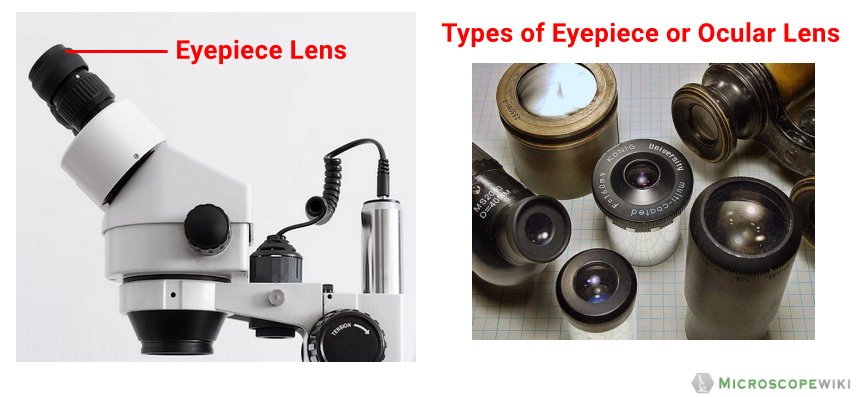
Eyepiece/Ocular
It is the lens used to magnify the main image produced by the objective. The eye peeps through the eyepiece to see the objects being visualized and studied.
Base
The purpose of the base is to support the entire microscope. It holds the microscope upright.
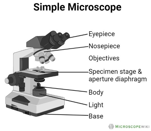
Image created with Biorender
Tube/Body Tube
It serves as the connector between the eyepiece/ocular and objective lenses.

Objective lenses
The lenses have varying magnifying power, which typically consists of 10x, 40x, and 100x. They have corresponding colors and they can be moved or rotated depending on your needs.

Revolving Nosepiece/Turret
It is used to hold the objective lenses in place. Through the revolving nosepiece or turret, you will be able to rotate the lenses making it easy for you to choose a lens that allow you to view the samples clearly.

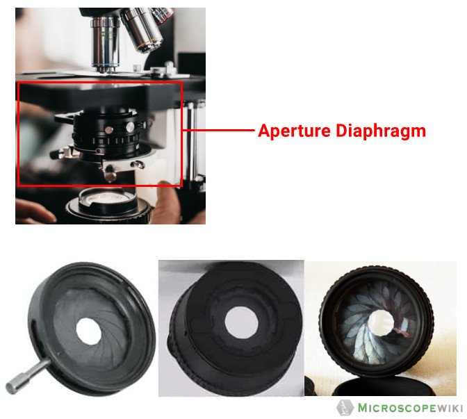
Diaphragm/Aperture Diaphragm
The diaphragm controls or regulates the amount of light that passes through the stage of the microscope.
Stage
It serves as the platform on which slides with samples are placed.

Stage Clips
These are utilized to fasten the slides in position.

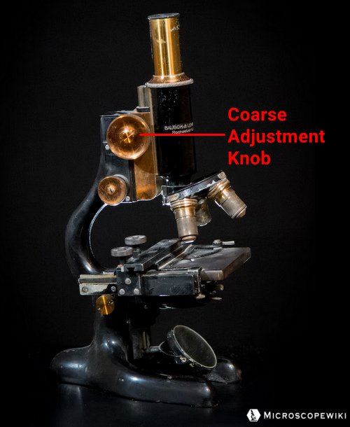
Coarse Adjustment Knob
Its purpose is to focus the scanning.
Fine Adjustment Knob
When looking at greater magnifications, a slow but exact control is employed to fine focus the image.

Arm
The arm of the microscope serves as the connector to the base. It also supports the tube. Although the arm in some microscopes is curved and in others is straight, all microscopes’ arm perform the same function.

Power switch
The microscope can be started or stopped using the main power switch.

Condenser
It makes use of 400X power lenses to concentrate light on the sample.

Parts of Compound Microscope (Labeled Pictures)
A compound microscope is a complicated assembly of various lenses that produces a significantly enlarged image of minute living things as well as other delicate characteristics of cells and tissues.
The parts of the compound microscope is categorized into two – the mechanical parts and the optical parts. It is also known as bright-field microscope because it enables the light to pass directly through the source of light through the two lenses.
Let us discuss the different parts of a compound microscope.
a. Mechanical Parts of a Compound Microscope
Foot or Base
The base is a U-shaped structure that bears all of the compound microscope weight.

Pillar
It projects vertically and the main function is to support the stage by resting on the base.

Arm
The arm, a robust and curved component, is used to handle the entire microscope.
Stage
It is the flat plate that is rectangular in shape and attached to the lower part of the arm. The specimen is set up on the stage so that the various aspects can be studied and examined. There is a hole in the middle of the stage through which light can enter.

Inclination Joint
It is a junction where the arm is attached to the pillar of the compound microscope. The inclination joint can be used to tilt the microscope.
Clips
Two clips link the stage’s upper portion to them. With the aid of the clips, the slide can be kept in place.
Diaphragm
- Below the stage is where the diaphragm is fixed in place.
- It regulates and modifies the amount of light that enters the microscope.
- The quality of the image may be affected in very small but significant ways based on the type and settings used on the diaphragm.
- The diaphragm’s main job is to alter the angular aperture of the light cone that is created after passing through the condenser.
- The cone light’s size is crucial since you won’t obtain the best image quality if it is not in alignment with the ideal numerical aperture of the objective lens currently in use.
There are different varieties of the diaphragm:
- Disc diaphragm – A disc diaphragm, which is less frequent, resembles somewhat like a spinning wheel with various aperture diameters. Need more light? Change it to the wide hole. Desire less light? Choose the smallest of the two holes.

- Aperture Iris diaphragm – The iris diaphragm is the most typical form of diaphragm. These are a little more complex and more expensive. Because it performs the same function as the iris in our eyes, the iris diaphragm is given the term “iris.” Your iris regulates the amount of light that reaches your eye’s cones and rods by expanding or contracting. Your iris slowly expands to take in more light, similar to how it does when you are out in the dark for a minute as opposed to 15 minutes.
- Field Diaphragm – This diaphragm is situated nearer to the microscope’s light source. The same principles apply here, but the amount of light and the size of the resulting image’s field of vision are controlled.

Nose piece/Revolving Nosepiece/Turret
It is a circular structure with three holds in which the objective lenses are attached. it is usually made from metal material and you can rotate it, especially when selecting the right lenses.
Body Tube
The body tube, which is a hollow and tubular structure, is part of the arm’s top portion on the microscope. The adjustment knobs can be used to raise and lower the body tube.
Adjustment Knobs
It is found below the stage to regulate the stage’s forward/reverse and side-to-side movement. There are two types of adjustment knobs. These are fine adjustment knob and coarse adjustment knob.
- Fine adjustment knob – The smaller knob is used to focus an object precisely and sharply. This knob can be used to focus precisely and brings the object into sharpest focus.
- Coarse adjustment knob – It is a sizable knob used to raise and lower the body tube to precisely focus on the thing being inspected.
b. Optical Parts of a Compound Microscope
Eyepiece lens or Ocular
- A lens, referred as the eyepiece, is mounted at the upper part of the body tube.
- With the aid of an eyepiece, the object’s enlarged picture can be seen.
- There are several markings, which include 5X, 10X, and 15x on the eyepiece’s rim.
- These show the magnification strength. It is the lens that is placed on the body tube’s upper portion and is used to observe the specimens.

Mirror
- The lower end of the arm or the pillar has a mirror fastened to it.
- On one side is a regular mirror, and on the other is a concave mirror. It is used to reflect light into the microscope for a sharper view of the specimen.
- A compound microscope primarily makes use of concave mirrors. Plane mirrors are occasionally also used.
- Concave mirrors are required for higher magnifications; plane mirrors can be used for lower magnification.
Objective Lenses
- The specimen’s image is magnified by the objective lens, which also projects the enlarged picture into the body tube.
- The most popular magnification powers for objective lenses are 4x (scanning), 10x (low power), 40x (high power), and 100x (oil immersion).
Scanning Objective Lens (4x)
- The smallest magnification offered by any objective lens is that of a scanning objective lens.
- A 4x scanning objective lens provides a total magnification of 40x when paired with a 10x eyepiece lens, which is a common magnification for scanning objectives.
- Because they give viewers roughly sufficient magnification for a good view of the slide, or a “scan,” of the slide.

Low Power Objective (10x)
- One of the most useful lenses for monitoring and evaluating glass slide samples is the low power objective lens.
- It has more magnification power when compared with the scanning objective lens.
- You can see the slide more clearly without the need to get too close.
- Thanks to the 100x total magnification that a low power objective lens and a 10x eyepiece lens provide.

High Power Objective Lens (40x)
- For observing minute features within a specimen sample, a high-powered objective lens, often known as a “high dry” lens, is perfect. You can see a very detailed image of the specimen on your slide.
- Thanks to the 400x total magnification that a high-power objective lens and a 10x eyepiece provide.

Oil Immersion Objective Lens (100x)
- The most potent magnification is provided by this type of objective lens, which, when used with a 10x eyepiece, offers a staggering total magnification of 1000x.
- However, because the refractive indices of air and your glass slide differ somewhat, a special immersion oil is required to fill the gap.
- The oil immersion objective lens won’t work properly, the specimen will look blurry, and you won’t get the best magnification or resolution if you do not add a drop of immersion oil.
- Some manufacturers also offer oil immersion lenses in lower magnifications, which offer better resolutions.

Specialty Objective Lenses
- There are a number of additional objective lens magnifications that are useful for specific applications. The 2x objective, which is frequently used in pathology, has just half the magnification of a 4x scanning lens, giving the sample on the slide a better overall view.
- The gold standard for examining blood smears is the 50x oil immersion objective, which is frequently substituted for the 40x objective.
- The 60x objective, which is frequently offered in dry or oil immersion, magnifies objects by 50% more than a 40x lens. For larger magnification without the use of immersion oil, the 60x dry lens is sometimes preferred over a 100x oil immersion lens.
- Finally, the 100x dry objective can give great magnification without the use of immersion oil.
- The numerical objecure of a 100x dry objective is substantially lower when compared to the100x oil immersion objective, and as a result, the lens’s capacity to resolve small features in the specimen is also much lower.

References
- http://bioweb.uwlax.edu/APlab/Lab-Unit-01/Lab-01-04.html
- https://idea-bio.com/how-oil-immersion-objectives-can-improve-your-microscopy/
- https://www.ncbi.nlm.nih.gov/pmc/articles/mid/NIHMS363450/
- https://opg.optica.org/josa/abstract.cfm?uri=josa-41-2-111
- https://www.nature.com/articles/s41598-018-22561-w
- https://www.wjoud.com/doi/WJOUD/pdf/10.5005/jp-journals-10015-1495
- https://www.ncbi.nlm.nih.gov/pmc/articles/mid/NIHMS502860/
- https://microbenotes.com/compound-microscope-principle-instrumentation-and-applications/
- https://www.scirp.org/journal/paperinformation.aspx?paperid=68176
- https://unacademy.com/content/neet-ug/study-material/physics/a-basic-study-on-simple-microscope/