What is Bright-field microscope
A bright-field microscope, also known as a compound light microscope is among the simplest of optical microscopes. Optical microscopes employ visible light and a series of lenses to magnify the specimen and view it in detail.
A bright-field microscope uses light rays to create a dark image against a bright background and it is this contrast that makes this microscope simple and effective for use.
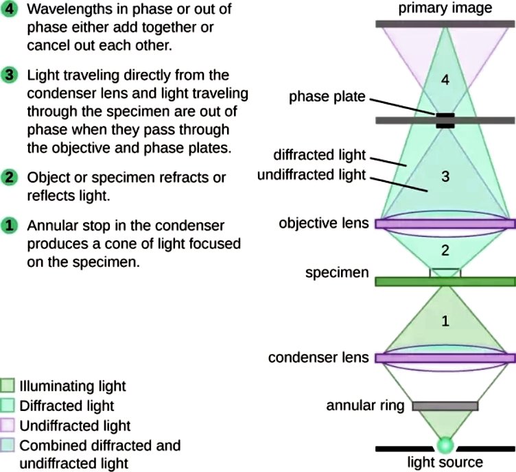
Working Principle and Diagram
Bright-field microscope works on the simple principle of absorption of light.
Firstly, the specimen is put on the stage and the light below is focused on the specimen and the specimen absorbs the light and the contrast image that is dark is viewed against a bright background thus creating an image that is magnified and viewed using the objective and ocular lens.
The specimens are usually stained with light absorbing materials such as nano gold particles to create a differential radiation pattern making it easier to distinguish the specimen against the bright background.

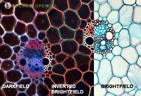
Difference between dark field and bright field illumination
Magnification and Resolution
- A bright-field microscope’s ability to create a high-resolution image from a well-illuminated light source that is focused on the specimen make it a simple yet effective microscope.
- The magnification level achieved by the bright-field microscope is dependent on the objective lens.
- A typical microscope has two to five lenses with varying degrees of magnification powers, and they create an enlarged image of the specimen which is further magnified by the ocular lens.
A bright-field microscope has a magnification powers of 40x- 1000x depending on the model of the microscope and the formula to calculate the power of a bright-field microscope is:
Total Magnification power = Objective lens magnification * Eyepiece magnification
The objective lens magnifies the specimen at a given resolution. The resolution is a very important aspect in the microscope, and it is the ability of the lens to distinguish clearly between the extremely tiny objects in the specimen and the ocular lens enlarge the high-resolution image created by the objective lens at the viewpoint.
Both the lenses together create a clear and magnified image of the specimen to allow for proper and accurate analysis.
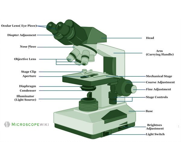
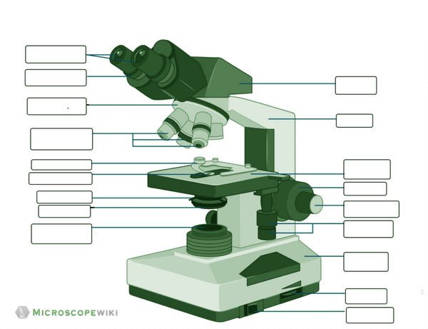
Parts of a bright-field microscope
A bright-field microscope is made up of the following parts
- Ocular Lens
- Objective Lens
- Focusing knobs
- Stage
- Condenser
- Arm
- Diaphragm
- Illuminating Source
- Nosepiece
Ocular Lens
This microscope has two eye lenses or ocular lens on the top of the microscope that are used to focus the image from the objective lens. It is from these lenses that we see the magnified image of the specimen.
Objective Lens
These are a set of lenses which make the image of the specimen larger and clearer, and this image is magnified using the ocular lens.
Focusing knobs
There are two knobs that help the user in focusing the lens on to the specimen. There is a coarse adjustment knob on the arm of the microscope that is used to move the nosepiece or the stage to focus on the image. There is also a fine adjustment that is used to make finer adjustment to ensure better image quality.
Stage
This is the place where the specimen is put, and it is below the objective lens. The stage is movable to allow for better focusing of the light on to the specimen.
Condenser
It is mounted just below the stage is used to focus the light beam on the specimen placed on the stage. The condenser is used to adjust the quality and intensity of light required. The power of the condenser is dependent on the model of the microscope.
Arm
This the backbone of the microscope and this metallic part is what on which the microscope rests and it is used to carry the microscope from one place to another. The arm is connected to the base and together both the base and the arm hold all the parts of the microscope.
Diaphragm
This is a very important part that allows for the adjustment of diameter of the light beam that passes through the condenser. When the diameter is very less, the light comes with high intensity thus creating a very high contrast image and conversely when the diaphragm is open the image is very bright with low contrast.
Illuminating Source
The microscope also has a mirror or an illuminator at the base of the microscope or at the nosepiece of the microscope
Nosepiece
This nosepiece holds about two to five objective lenses that have different magnifying powers. The nosepiece can be moved round to any position as per requirement of the objective lenses to focus on the specimen
Applications and Uses
The simplicity of the bright-field microscope makes it an ideal magnification device in many fields including microbiology, parasitology, bacteriology, and biology.
The most important thing, however, is that it is better used with stained specimens or specimens that can be stained using external materials. A few applications of the bright-field microscope include:
- Used to observe, analyze, and study plant cells
- Used to view, magnify, and study about animal cells
- Used to clearly study the morphologies of bacterial, and viral organisms
- Also used in the study of parasites like paramecium
- It finds use in agricultural laboratories to study soil samples and microorganisms in the soil
- It is also used in forensic labs and also diagnostic labs to study the various aspects of blood
- It is also used in studying various fungal cells that infest plants, animals, and human beings as well.
Advantages
- It is very easy to use with minimal requirements needed to operate and adjust the settings of the microscope while viewing the image
- The optics of the bright-field microscope don’t change the color of the specimen allowing for an accurate analysis
- It can be used for both unstained and stained specimens
- It is economical and is also comparatively light weight for use in various places like schools, labs etc.
- This microscope can be modified, and it allows for installing a digital camera to facilitate for better viewing making it a digital microscope
Disadvantages
- It can’t be used to view live samples and only fixed specimens can be viewed under a bright-field microscope
- The typical configuration has only 100x magnification range which is too low. But modification of the microscope allows for magnification up to 1000x
- The diaphragm may cause very high contrast thereby rendering a distortion in the image and causing a poor analysis. So, an iris diaphragm is preferred.
- The typically low contrast produced by the microscope due the poor absorption of light by the specimen requires the user to resort to staining to obtain better results
- The staining may result in extraneous substances creating unwanted details in the image of the specimen
Frequently Asked Questions
Q1. What can influence the resolution of a bright field microscope?
Magnification, wavelength of light and quality of lens are the three aspects that can affect the resolution of the bright-field microscope
Q2. Can you view live organisms with a bright field microscope?
No, the bright-field microscope can be used to observe only fixed specimens placed on the stage of the microscope. However, with the latest advances, it is still possible to view them, but modifications need to be made.
Q3. Do both bright field and dark field microscopes have condenser?
Yes, both the microscopes have condensers, but they are used differently to obtain the desired image.
Q4. Does bright field microscope look at live or dead?
The bright-field microscope finds more usage at observing fixed (dead) specimens as the specimens need to absorb the light and create a contrast image against a bright background. However, certain modifications and technology advances have made the observation of live organisms a reality.
Q5. Is bright field microscope reflection or refraction?
It creates and image by focusing light from the illuminator and this light is differentially, absorbed, transmitted, refracted, or reflected by different structures.
Q6. Should I buy a bright field microscope?
Depending on the nature of work, one can buy a bright-field microscope. It finds extensive usage in the fields of biology, parasitology, microbiology, forensics etc.
Q7. What controls illumination in bright field microscope?
The condenser in the bright-field microscope houses an aperture diaphragm that controls the intensity of light to be focused on the specimen
References:
- https://www.ruf.rice.edu/~bioslabs/methods/microscopy/microscopy.html
- https://microscopeclarity.com/bright-field-microscopy/
- https://www.geog.ucl.ac.uk/resources/laboratory/light-microscopy/bright-field-microscopy
- https://www.microscopemaster.com/brightfield-microscopy.html
- https://microbenotes.com/brightfield-microscope/
- https://www.sciencedirect.com/topics/medicine-and-dentistry/bright-field-microscopy
- https://en.wikipedia.org/wiki/Bright-field_microscopy
- https://www.microscopemaster.com/microscope-resolution.html